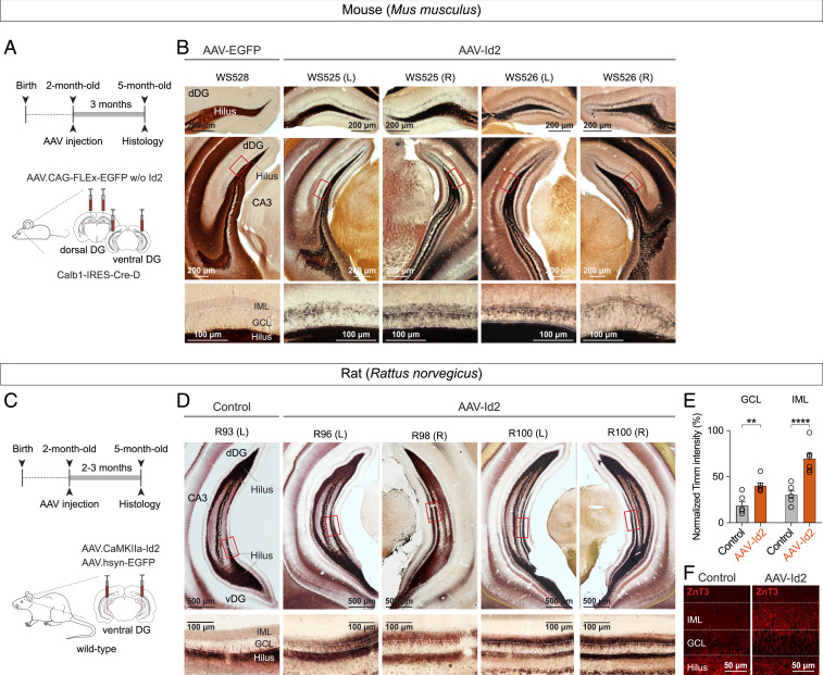
Fig. 3.
AAV-delivered Id2 induces MF rewiring throughout the mouse and rat hippocampus. (A) Experimental design showing Id2 overexpression in mouse dorsal and ventral hippocampus. (B) Images show Timm's-stained sections collected from different levels of dorsal hippocampus (bregma, –2.0 mm and –3.2 mm) after AAV-EGFP (control) and AAV-Id2 injections. Higher-magnification images at bottom show sprouting in GCL and IML in AAV-Id2 mice. (C) Experimental design showing Id2 overexpression in rat ventral hippocampus. (D) Example images of Timm staining in rats after Id2 overexpression. Coronal sections of rat ventral hippocampus (bregma, –6.2 mm) were collected from regions where AAV infection was confirmed by EGFP expression. Non-AAV-infected hippocampus was used as control. (E) Quantification of Timm’s staining intensity. Intensities were measured relative to signals in the hilus of the same sections (two-way ANOVA, GCL: Control versus AAV-Id2, **P = 0.0017; IML: Control versus AAV-Id2, ****P < 0.0001). (F) ZnT3 staining of MF synapses in GCL/IML 3 mo after AAV-Id2 injections.

