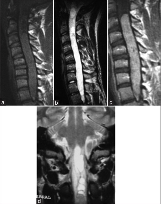
Figure 2.

Spinal MRI: A case of Cervico Dorsal Ependymoma. Sagittal section on T1 sequence (a), T2 sequence (b), and T1 after gadolinium enhancement (c); Coronal section on T2 sequence: A tumoral process is centro medullar extended from C3 to D1 (d). This process has an isosignal on T1, it is hyperintense on t2 surrounded by a discreet T2 hypointense line. Enahancement is homogenous. There is an association with syringomyelic cavities and even syringobulbia
