
Figure 3.
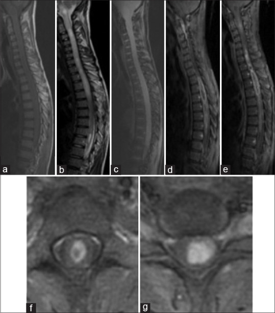
Spinal MRI: A case of Cervico Dorsal Astrocytoma, Sagittal section on T1 sequence (a), T2 sequence (b), gradient echo sequence (c) and T1 Fatsat after gadolinium enhancement (d and e); Axial sections on T1 Fatsat after Gadolinium enhancement (f,g): There is a tumoral process extended on all of the cervical and dorsal spinal cord, it has an isosignal on T1 and has a heterogenous hypersignal on T2. Enhancement is heterogenous and peripheral delimiting necrosis. It has multiple cysts with a syringomyelia extended to the bulb