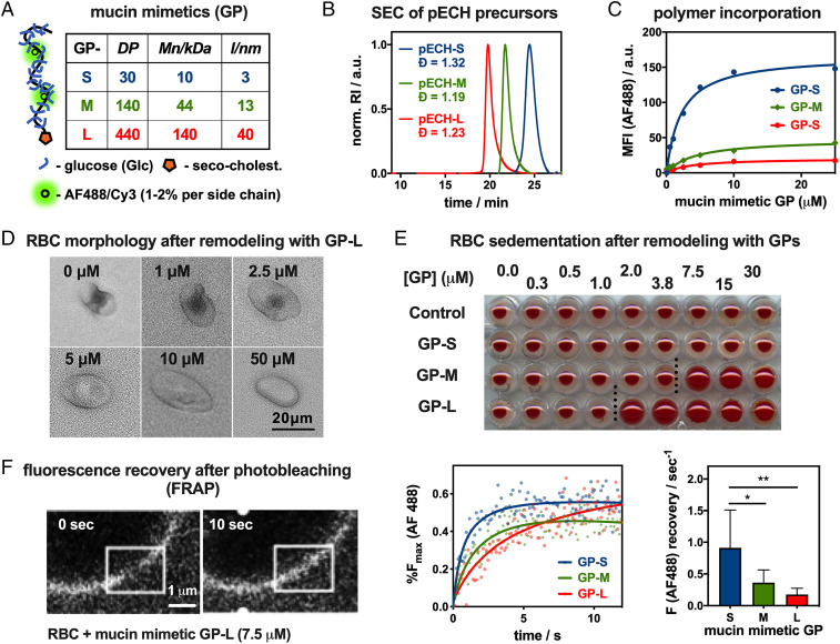
Fig. 2.
Construction and characterization of a mucosal glycocalyx model. (A) Short (S), medium (M), and long (L) mucin mimetic GPs ranging in size from ∼3 nm to 40 nm were generated from poly(epichlorohydrin) (pECH) precursors (Mn, number average molecular weight; l, estimated end to end length). (B) Size-exclusion chromatography (SEC) of pECH precursors indicates narrow molecular weight distributions of the polymer scaffolds. The polymers were modified with biologically inert glucose side chains, fluorescent probes (AF488 or Cy3) for visualization, and chain end-terminated with hydrophobic cholestenone membrane anchor. RI = refractive index. (C) Incorporation of AF488-labeled mucin mimetics GPs into RBC membranes was analyzed by flow cytometry. MFI = median fluorescence intensity. (D) Optical microscopy and light scattering reveal increased cell rounding with increasing glycocalyx density (shown for GP-L). (E) Sedimentation of RBCs remodeled with increasing concentration of all three GPs (cGP = 0.25 μM to 30 μM). (F) FRAP analysis shows length-dependent diffusion of AF488-labeled mucin mimetics GP in RBC membranes. White overlaid boxes draw attention to the photobleached region. Lines represent the average signal from n = 6 cells, cGP = 7.5 μM; P values were determined by one-way ANOVA with multiple comparisons (*<0.05, **<0.01).

