Fig. 4.
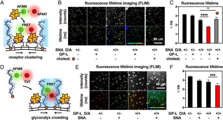
Glycocalyx crowding drives clustering of lectin-receptor adhesion complexes. (A) Changes in SNA clustering in response to glycocalyx crowding were assessed based on FRET between lectins labeled with donor (D = AF595) and acceptor (A = AF647) probes. (B) Representative FLIM images and (C) bar graph representations for the binding of SNA probes to RBCs before and after remodeling with GP-L (cGP = 7.5 μM) or the hydrophobic anchor S.5 alone (cS.5 = 7.5 μM). Decrease in fluorescence lifetime (τ) indicates closer SNA proximity. (D) Mucin mimetics labeled with donor (D = AF488) and acceptor (A = Cy3) probes were employed to measure changes in glycocalyx crowding after SNA binding via FRET. (E) Representative FLIM images and (F) bar graph representations of RBCs remodeled with mucin mimetic probes (cGP-D/A = 15 μM) before and after incubation with SNA. Decrease in fluorescence lifetime (τ) indicates enhanced glycocalyx crowding. Color scales represent fluorescence lifetime (τ/ns). Values represent averages and SDs for representative image frames containing >10 cells; P values were determined by student test; ***<0.001, ****<0.0001.

