Fig. 3.
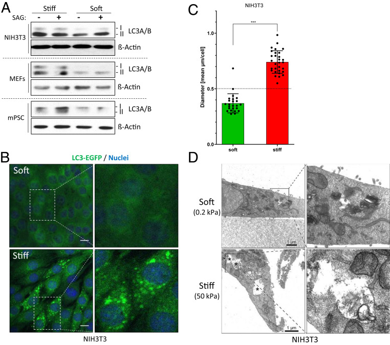
Substrate stiffness is sufficient to induce autophagy. (A) Levels of the autophagy marker LC3A/B in various cell lines as determined by Western blotting. Shown is one representative of n = 1 to 3 independent experiments. (B) Confocal images of NIH 3T3 cells stably expressing LC3B-EGFP (green) and grown in soft or stiff conditions. Nuclei appear in blue (DAPI). (Scale bar, 10 µm.) (C) Mean diameter of LC3B-EGFP spots per cell (as shown in B). Each dot represents one cell. (D) TEM images of NIH 3T3 cells cultured on soft or stiff hydrogels. APLs are indicated by asterisks. (Scale bar, 1 µm.)

