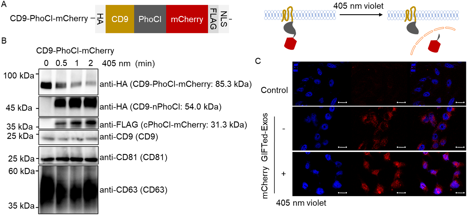
Figure 3.

Design, generation and examination of mCherry GIFTed-Exos. (A) Schematic of the design of mCherry GIFTed-Exos. (B) Immunoblot analysis of the mCherry GIFTed-Exos and violet light-induced release of mCherry. Theoretical molecular weights of fusion proteins are shown. (C) Cellular uptake of mCherry GIFTed-Exos. The exosomes (500 μg mL−1) without and with 405 nm violet light irradiation were incubated with HeLa cells for 6 hours at 37°C, followed by washing with PBS, permeabilization, and confocal imaging. Blue: nuclei stained with DAPI; red: mCherry. Scale bars, 20 μm.
