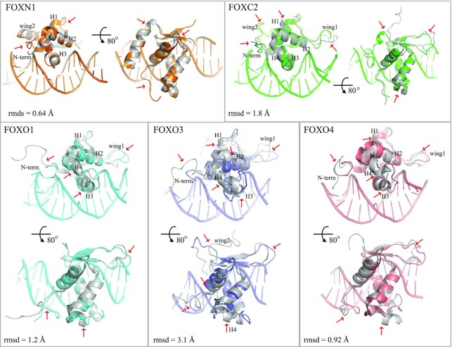
Figure 5.
DNA-binding-induced conformational change of FOX-DBD. DNA-bound structures are shown in color and apo FOX-DBD structures are shown in gray. Major conformational differences are indicated by red arrows. The PDB codes are FOXN1, 6EL8 (DNA-bound) and 5OCN (apo); FOXC2, 6AKO (DNA-bound) and 1D5V (apo); FOXO1, 3CO7 (DNA-bound) and 6QVW (apo); FOXO3, 2UZK (DNA-bound) and 2K86 (apo); FOXO4, 3L2C (DNA-bound) and 1E17 (apo).

