Figure 1.
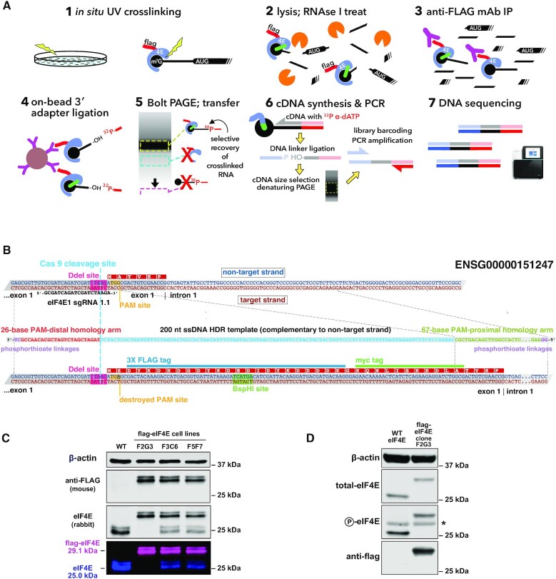
capCLIP method and flag-eIF4E expressing cell line. (A) Individual steps of the capCLIP method: (1) short-wavelength (∼254 nm) UV irradiation of living HeLa cells (clone F2G3; see below) expressing flag-eIF4E creates a ‘zero-order’ photo-cross-link between an mRNA’s m7G cap and an associated protein, e.g. eIF4E; (2) cells are then lysed and the mRNA fragmented using a limiting concentration of RNAse I; (3) flag-eIF4E (and cross-linked m7G-capped mRNA fragments) are captured by anti-FLAG mAb complexed to protein G-coated magnetic beads; (4) an RNA adapter is ligated ‘on bead’ to the 3′ end of the capped RNA fragment; (5) IP material is then subjected to neutral pH PAGE and then transferred to a membrane to size-separate cross-linked flag-eIF4E:mRNA:adapter complexes from unwanted IP products; (6) flag-eIF4E is proteolytically removed from capped mRNA:adapter complexes and the purified material used for cDNA synthesis, followed by ligation of a 3′-end DNA adapter and then PCR amplification; and (7) finally, barcoded capCLIP libraries are combined for high-throughput sequencing. (B) Outline of the CRISPR/Cas9-mediated addition of a 3X-flag/1X-myc epitope tag to the N-terminus of human eIF4E (Ensembl gene ENSG00000151247) in HeLa cells (henceforth identified as ‘flag-eIF4E’). Experimental details of this eIF4E gene editing strategy are provided in the ‘Materials and Methods’ section. (C) Anti-eIF4E/anti-flag westerns of wild-type (WT) HeLa cells and three CRISPR-edited flag-eIF4E HeLa clonal cell lines. The 29-kDa flag-eIF4E and the 25-kDa WT eIF4E bands detected by the rabbit anti-eIF4E antibody, and the 29-kDa flag-eIF4E band detected solely by the anti-flag antibody, are shown both as separate and as merged LiCor image channels. (D) Westerns using antibodies for total eIF4E, P-eIF4E or flag of lysates from WT and the 3X-flag-eIF4E-expressing HeLa cell clonal line F2G3. The asterisk denotes a non-specific band consistently seen with this anti-P-eIF4E antibody.

