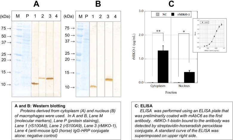
Fig. 5.
Detection of rMIKO-1 in the cytoplasm and nucleus of macrophages by Western blotting and ELISA. Western blotting was performed to detect rMIKO-1-biotin in the cytoplasm and nucleus of macrophages treated with rMIKO-1 at 37 C for 2 h in 5% CO2. (A) and (B) Lanes M, P, 1, 2, 3, and 4 indicate molecular markers, protein staining, rS100A8, rS100A9, rMIKO-1-biotin, and the negative control, respectively. rS100A8 and rS100A9 were stained using mAb8A6-HRP and mAb1D11-HRP conjugates, respectively. rMIKO-1-biotin was detected using the STA-HRP conjugate adequately diluted with a working solution of Blocking One (Nacalai Tesque, Kyoto, Japan), in which 3,3′-diaminobenzidine tetrahydrochloride n-hydrate and hydrogen peroxide were used as the substrates for color development. (C) Preparative ELISA for rMIKO-1 was performed to measure rMIKO-1-biotin using an ELISA plate that was coated with a monoclonal antibody of mAbC6 (5 μg/ml) as the first antibody. rMIKO-1-biotin bound to the first antibody was detected by the STA-HRP conjugate. HRP activity was spectrophotometrically assessed. A standard curve for rMIKO-1-biotin was superimposed on the upper right side. Error bars are shown as means±SD values. *, P<0.05; **, P<0.01.

