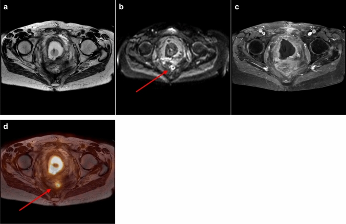
Fig. 4.
a–d Small presacral tumor recurrence. a–c Axial T2-weighted/axial diffusion-weighted/axial T1-weighted contrast-enhanced MRI. MRI showing diffuse T2-hypointense changes presacrally, a small rim-like diffusion restriction and no focal contrast enhancement. Diffusion restriction was thought to be due to reactive/inflammatory changes. MRI was scored as recurrence unlikely (score 1). d Corresponding axial PET/MRI fusion image demonstrating a small presacral lesion with intense FDG-uptake, which was interpreted as highly suspicious (score 4) for tumor recurrence. Biopsy confirmed tumor recurrence

