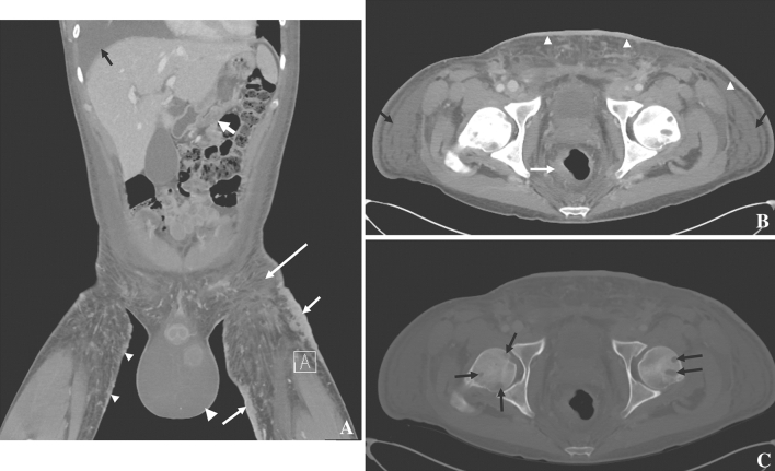
Fig. 4.
33-year-old male with HIV and KS. A Coronal contrast-enhanced CT showing antral wall thickening (fat white arrow), large and thick skin thickening (plaques, short white arrows), small skin nodules (small arrowheads), hydroceles (large arrowhead), subcutaneous edema (long arrow), and a pleural effusion (black arrow). B Axial contrast-enhanced CT showing asymmetric rectal wall thickening (white arrow), skin thickening (arrowheads), and subcutaneous edema (black arrows); C bone windows show lytic bone metastases

