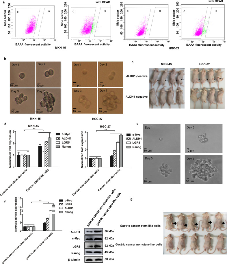
Fig. 1. Sorting of gastric cancer stem-like cells.
a Sorting of gastric cancer stem-like cells. The fluorescence activated cell sorting was performed based on the detection of ALDH1 activity using the ALDH1 fluorescent substrate BODIPY-aminoacate (BAAA). As a control, the activity of ALDH1 was inhibited by DEAB. The ALDH1-positive cells were potential gastric cancer stem-like cells (D region) and ALDH1-negative cells were cancer non-stem-like cells (C region). b Tumorsphere formation assay. The ALDH1-positive cells were subjected to tumorsphere formation assay. The sphere formation was examined with a light microscope. Scale bar, 100 μm. c Tumorigenicity of cancer stem-like cells in nude mice. Mice were subcutaneously injected with ALDH1-positive or ALDH1-negative cells. Forty days later, the tumors were examined. Arrows indicate the tumors. d Differential expressions of stemness-associated genes in gastric cancer stem-like cells and non-stem-like cells. Quantitative real-time PCR was conducted to detect the mRNA levels (**p < 0.01). e Tumorsphere formation of gastric cancer stem-like cells from the solid tumors of patients with gastric cancer. Scale bar, 10 μm. f The expression levels of stemness genes in gastric cancer stem-like cells isolated from the solid tumors of gastric cancer patients. The statistical significance of difference between treatments was indicated with asterisks (**p < 0.01). g Tumorigenicity of the potential gastric cancer stem-like cells in mice. Nude mice were subcutaneously injected with the potential gastric cancer stem-like cells or gastric cancer non-stem-like cells. Forty days later, the tumors were examined. Arrows indicate the tumors.

