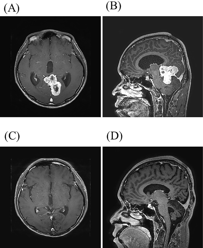
Figure 1.
(A, B) Magnetic resonance imaging (MRI) before surgery. Axial (A) and sagittal (B) contrast-enhanced T1-weighted images reveal a heterogeneously enhanced tumor in the left occipital lobe and cerebellum. (C, D) Follow-up MRI after surgery and radiation therapy. Axial (C) and sagittal (D) contrast-enhanced T1-weighted images showed no evidence of a residual lesion.

