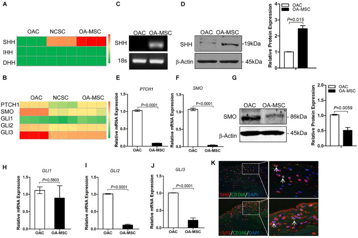
FIGURE 1.
Sonic Hedgehog (SHH) was synthesized by human cartilage mesenchymal stromal cell (MSC) while hedgehog (HH) receptors and transcription factors were expressed by human chondrocyte. (A) Heat map of copy numbers of transcripts encoding HH ligands (SHH, IHH, DHH) in osteoarthritic chondrocytes (OAC), normal cartilage stromal cells (NCSC), and osteoarthritis mesenchymal stromal cell (OA-MSC). (B) Heat map of copy numbers of transcripts encoding HH receptors (PTCH1, SMO) and transcription factors (GLI1, GLI2, GLI3). (C) Real-time RT-PCR analysis validated the upregulation of SHH transcript in OA-MSC. Total RNA was isolated from primary human OAC and OA-MSC. 18S RNA was used as RT-PCR and loading control. Data are representative of three independent experiments. (D) Western blot analysis and quantification indicated SHH protein was abundantly expressed by OA-MSC but not OAC. β-actin was used as Western blot analysis and protein loading control. Molecular weight of proteins was indicated. Relative levels of SHH protein expression in OA-MSC are shown in the bar graph (n = 3). Real-time RT-PCR analysis validated the up-regulation of PTCH1 transcript (E) and SMO transcript (F) in OAC. Total RNA was isolated from primary human OAC and OA-MSC. 18S RNA was used as a normalizing control (p < 0.0001). (G) Western blot analysis and quantification indicated SMO protein was down-regulated in OA-MSC. β-actin was used as Western blot analysis and protein loading control. Molecular weight of proteins is indicated. Relative levels of SHH protein expression in OA-MSC are shown in the bar graph (n ≥ 4). Real-time RT-PCR analysis validated the expression of GLI1 transcript (H), GLI2 transcript (I), and GLI3 transcript (J) in OAC and OA-MSC. Total RNA was isolated from primary human OAC and OA-MSC. 18S RNA was used as a normalizing control (n ≥ 4). (K) Double immunofluorescence histochemical analysis of human osteoarthritis (OA) articular cartilage with anti-sonic hedgehog antibody (rhodamine red), anti-CD166 (cartilage MSC marker) antibody (fluorescein green), and Hoechst nuclei dye (DAPI blue). SHH protein was distributed in CD166-positive OA-MSC cells which synthesized SHH (arrows). SHH protein was also distributed in CD166-negative OACs which SHH bound. SMO protein was distributed in CD166-negative chondrocytes (arrows). The images shown are representative of multiple tissue samples (n = 3). Scale bar = 125 μm.

