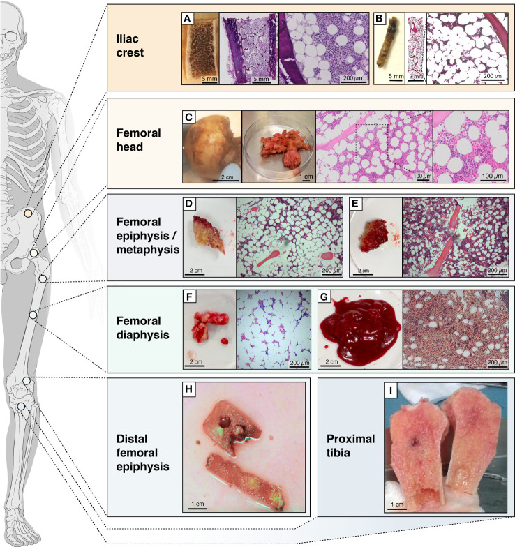
Figure 2.
Schematic figure showing the locations from which BMAT is obtained. Locations from which BMAT is obtained, heterogeneity in tissues (e.g. more fatty vs less fatty) and collected fractions. (A) transiliac bone autopsy obtained with a Bordier trephine and (B) iliac bone marrow biopsies obtained with a Jamshidi trephine; (C) femoral head autopsy; (D) epiphyseal or (E) metaphyseal tissue from femur; (F) diaphyseal bone or (G) bone marrow from femur; (H) tissue from the distal femoral epiphysis; (I) bone tissue from the proximal tibia.

