Figure 5.
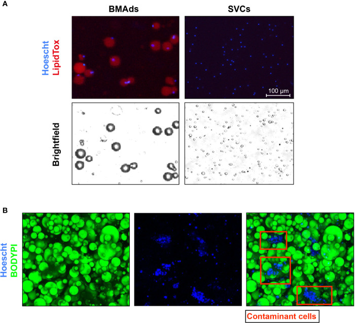
Histological images demonstrating the purity of the BMAds. (A) BMAds were stained with Hoechst and LipidTox DeepRed. Stromal-vascular cells (SVCs) were included as a negative control for LipidTox staining. Stained cells were then analysed using a Nexcelom Vision Cellometer. Adipocyte images confirm that all nuclei are associated with unilocular lipid droplets and there are no lipid-free nuclei, suggesting an absence of contaminating non-adipose cell types. Analysis of the SVC fraction confirms that the LipidTox signal is dependent on the presence of lipid droplets. Scale bar represents 100 μm. (B) BMAds were stained with BODIPY and Topro 3 to verify by IF the integrity of BMAds (BODIPY staining) and the purity of the adipocyte suspension. Nucleus staining show the presence of contaminant cells attached to adipocytes (red squares).

