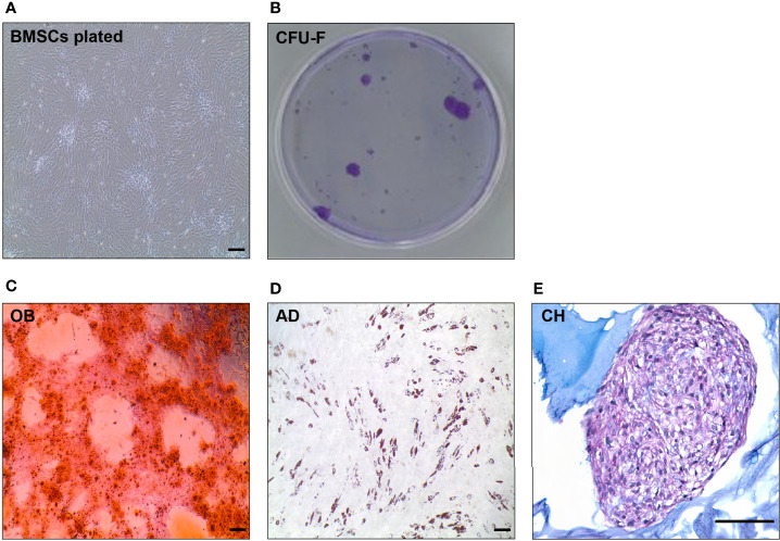
Figure 7.
In vitro evaluation of BMSC quality (BMSC culture, CFU-C, tri-differentiation). (A) Representative picture of spindle-like shape cells in in vitro culture of human BMSCs. (B) CFU-F of BMSCs after 14 days in vitro culture visualized by Violet Blue in a petri dish. (C) Alizarin staining of mineralized matrix in 10 days osteoblast (OB) culture differentiated from primary BMSCs. (D) Oil Red O staining of neutral lipids in a 10 days adipocyte (AD) culture differentiated from primary BMSCs. (E) Alcian Blue staining of a 21 days chondrocyte (CH) culture differentiated from BMSCs. Scale bar represents 200 μm in all the pictures.

