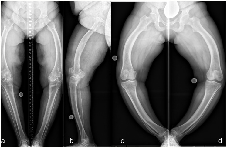
Figure 6.
Lower limb deformity of patients prior to surgical frontal plane correction at our hospital. Left (A frontal, B sagittal): 50 y/o, female, five prior surgeries, GDI: 43.7, BMI: 28.7 kg/m2, conventional oral medication. Right: 33 y/o, female, no previous surgeries prior to presentation, GDI: 27.6, BMI: 37.6 kg/m2. Full-length anteroposterior radiograph (hip to ankle) of both lower limbs in standing position was not performed due to severe varus deformity. Thus, radiograph image of each leg was taken individually (C, D).

