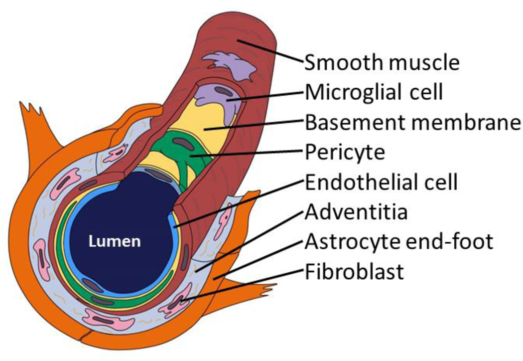
Fig. 1. A brain arteriole and constituents of the cerebral arteriolar vascular unit (cAVU).
Brain arterioles are not all alike, but this schematic depiction may orient readers to some cAVU components. Shown here is a relatively thin-walled arteriole--no internal elastic lamina or adventitial nerve fibers are depicted. Inflammatory cells including macrophages and microglia are commonly found in and around the arteriolar wall. Acellular strands of elastin and collagen are interwoven with fibroblast-like cells in the adventitia.

