
Fig. 4. Arteriolar walls can show different histomorphologies with aging.
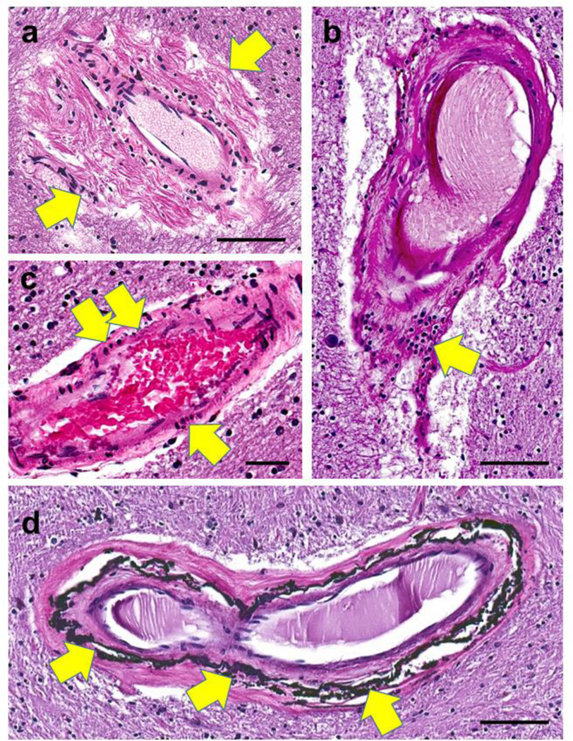
In panel (a), periarteriolar (adventitial) fibrosis is extensive and non-concentric (arrows). Panel (b) shows a collection of lymphocytes (arrow) in portions of the vessel wall. In panel (c), vessel wall changes include pyknotic-appearing smooth muscle cells (arrows). Siderocalcinosis, distinct from B-ASC, has been associated with dementia [194] and is usually seen preferentially in the globus pallidus (arrows in d). Synonyms for this include medial vascular calcification, calcific medial arteriosclerosis, and Monckeberg’s medial sclerosis. Scale bars = (a, b, and d): 100 μm; (c): 60 μm.