
Figure 1.
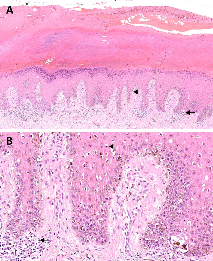
Acral melanoma. (A) In situ acral lentiginous melanoma with a lentiginous proliferation of atypical melanocytes along the dermoepidermal junction (arrowhead), with pagetoid spread and scattered nests at the tips of the rete ridges (arrow). There is also acanthosis, hypergranulosis, and hyperkeratosis (H&E, ×100). (B) Tumour cells are hyperchromatic and have a cytoplasmic fixation retraction artefact (arrowhead). The dermis contains a chronic inflammatory cell infiltrate associated with macrophages (arrow) (H&E, ×400).