
Figure 2.
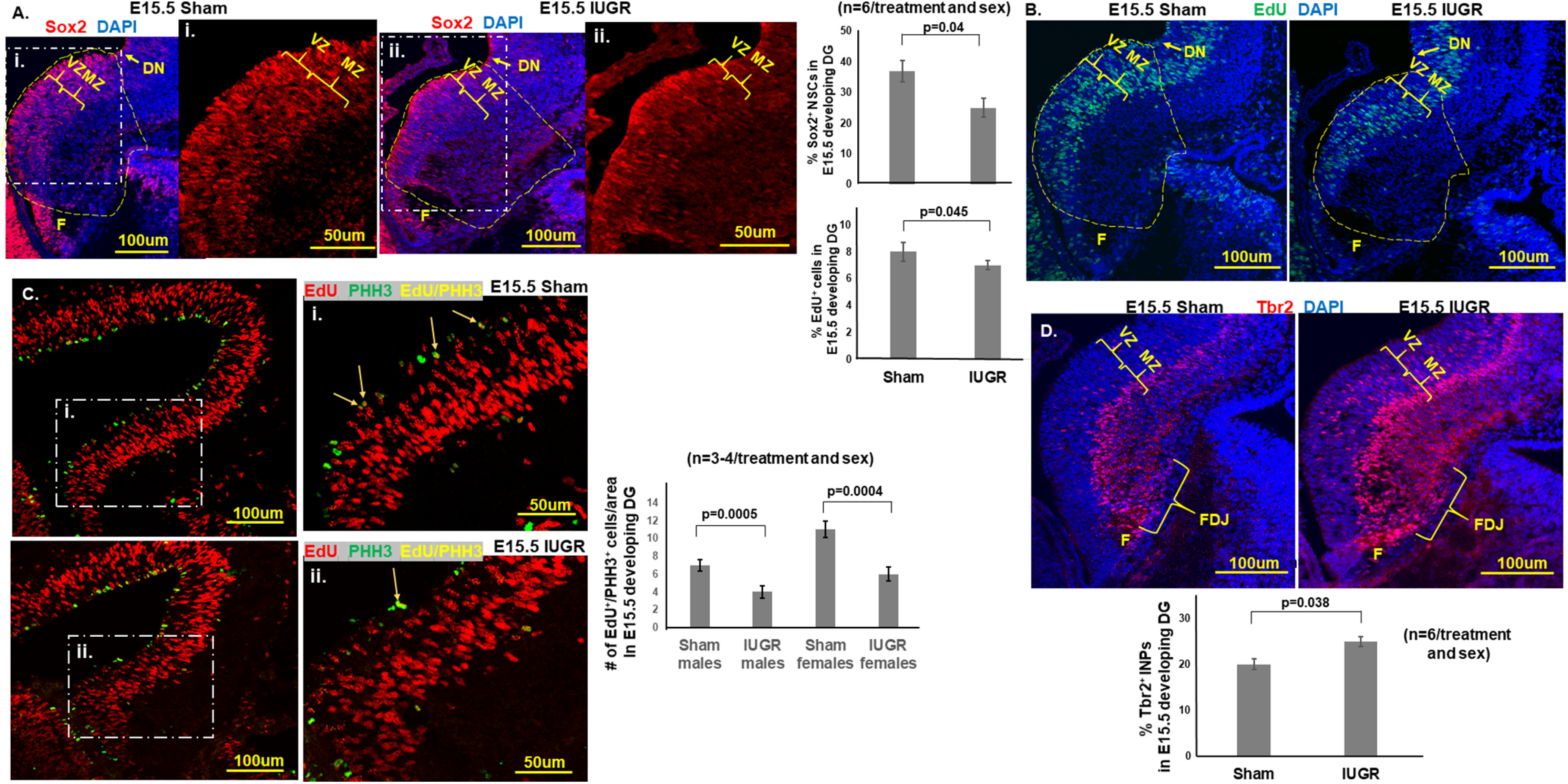
A, Representative photomicrographs of Sox2+ NSCs in E15.5 developing DG. Blue DAPI stain denoted cell nuclei. i, ii, Magnified views of Sox2+ NSCs in the white dashed regions of A. IUGR offspring of both sexes showed decreased percentages of Sox2+ NSCs (number of Sox2+ cells divided by the number of DAPI+ cells in the yellow dashed outlined area) in the VZ and MZ. p = 0.04, IUGR males and females compared with sex-matched shams. F, Fimbria. Yellow dashed outlines delineate the quantified areas that were manually traced in ImageJ beginning at the dentate notch (DN) to the beginning of the fimbria on the ventricular side and across to the pial surface. B, Representative photomicrographs of EdU+ proliferative cells in E15.5 developing DG. Blue DAPI stain denoted cell nuclei. IUGR offspring of both sexes showed decreased percentages of EdU+ proliferative cells (the number of EdU+ cells divided by the number of DAPI+ cells in the yellow dashed outlined area; i.e., cells in S phase of cell cycle, in VZ and MZ. p = 0.045, IUGR males and females compared with sex-matched sham controls. Student’s t test was used to compare sham and IUGR offspring for Sox2 and EdU, given that no differences were noted when sex was separated. C, Representative photomicrographs of E15.5 sham and IUGR developing DG with EdU and pHH3 at Ser 10 immunofluorescent costaining. White dashed boxed areas denoted as i and ii are enlarged regions demonstrating the colocalization of red EdU+ cells with green pHH3+ cells in yellow (arrows). Both IUGR males (p = 0005) and females (p = 0.0004) had a decreased number of EdU+/PHH3+ colocalized cells compared with sex-matched shams showing that IUGR progenitors had fewer cells completing mitosis of the cell cycle. Two-way ANOVA with Tukey’s HSD post hoc test was used to examine the effects of treatment (sham or IUGR), sex, or both sex and treatment on the number of EdU+/pHH3+ colocalized cells. D, Representative photomicrographs of Tbr2+ in E15.5 developing DG. Blue DAPI stain denoted cell nuclei. IUGR offspring of both sexes showed increased percentages of Tbr2+ INPs (the number of Tbr2+ cells divided by the number of DAPI+ cells) in the VZ, MZ, and FDJ. p = 0.038, IUGR males and females compared with sex-matched sham controls. Student’s t test was used to compare sham and IUGR offspring given no differences were noted when sex was separated.