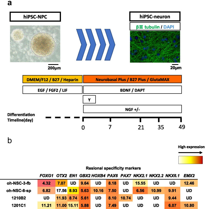
Fig. 1.
Procedure for the neuronal differentiation and characterization of hiPSC-NPCs. a Procedure for the neuronal differentiation of hiPSC-NPCs. The phase-contrast image shows the neurosphere of the hiPSC-NPCs before differentiation, and the immunofluorescence image shows neurons derived from hiPSC-NPCs (green: βIII-tubulin, blue: DAPI). Scale bars: 200 μm and 20 μm. b Gene expression of hiPSC-NPCs (1210B2 and 1201C1) and human neural tissue-derived NSPCs (oh-NSC-3-fb and oh-NSC-8-sp) before differentiation. The ΔCt values are shown. Red: high expression. UD undetermined

