Figure.
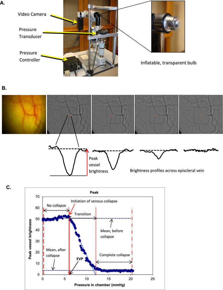
Measuring episcleral venous pressure. (A) Episcleral venomanometer. An episcleral venomanometer mounted on a slit lamp biomicroscope and modified for automated control. (B) Recording of venous collapse. The venous collapse was recorded by using a video camera. Brightness profiles across a selected segment of the vein were calculated using custom image analysis software. (C) Change in brightness profile across an episcleral vein. The peak brightness across the vein was graphed as a function of applied pressure.

