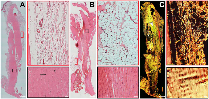
Figure 1.
Histological structure of healthy Achilles tendon. Representative serial longitudinal sections of Achilles tendons, with two fields of view of paratenon (red boxes) and a region of the mid tendon section (black boxes). (A) Formalin-fixed, hematoxylin–eosin (H&E)-stained section shows uniformly organized parallel collagen bundles and elongated tenocyte cell nuclei (black arrows). The paratenon is seen as loose connective tissue surrounding the tendon. (B) Bouin-fixed, H&E-stained tendon shows collagen fibers appearing intensely eosinophilic with nuclei stained darkly. In addition, the shrinkage effect of picric acid in Bouin solution causes artifactual changes in the normal appearance of tendon. (C) Formalin-fixed, picrosirius red staining on a sequential tissue section from the same tendon as panel A shows thick, bright yellowish-orange fibers. Another tendon area with greenish collagen fibers (white arrowhead) also contains some fibers that are at extinction (white arrows). The paratenon is seen with both thin fibers and thick fibers of yellowish-orange birefringence by polarizing microscopy. Tendon overview images were obtained with a 4× magnification lens (scale bar: 500 µm). Higher magnification images within the boxes were obtained with a 20× magnification lens (scale bar: 50 µm).

