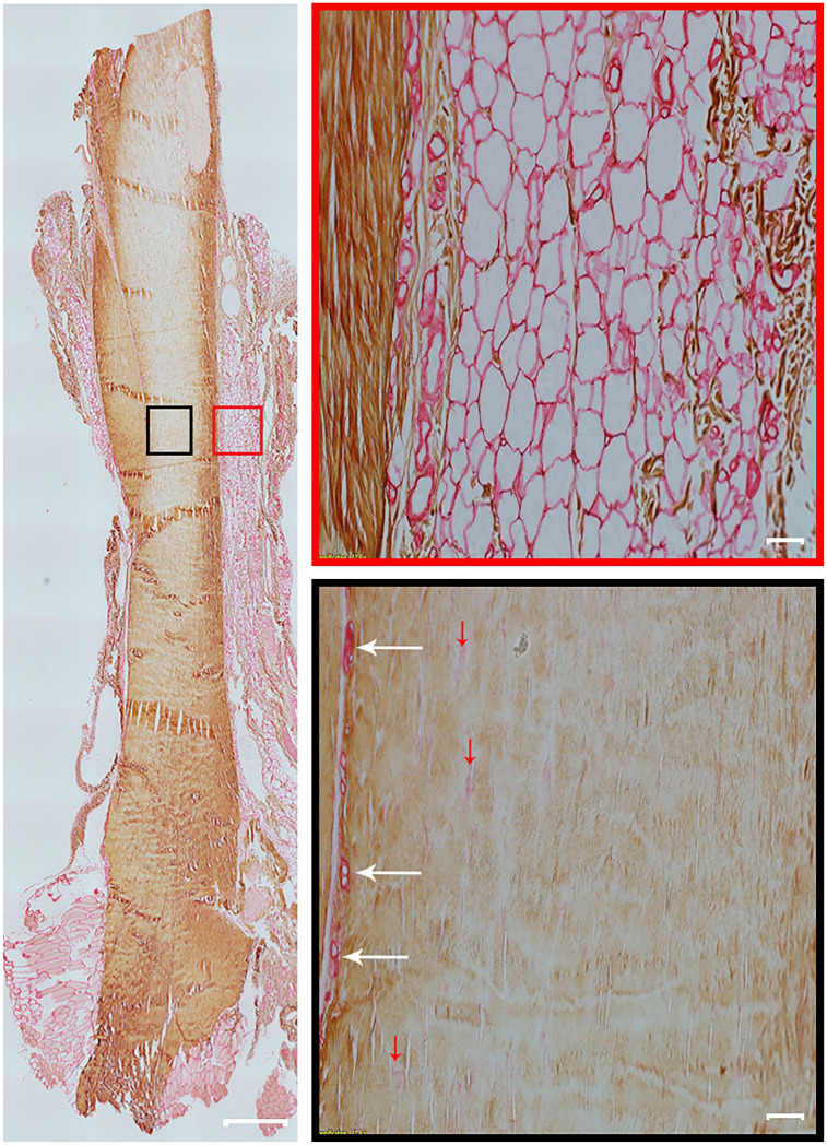
Figure 4.
Collagen type I and type III expression in rat Achilles tendon. A representative entire longitudinal Achilles tendon section is double immunolabeled for collagen type I and collagen type III. Positive immunolabelling for collagen type I is brown (diaminobenzidine chromogen) and for collagen type III is red (permanent red chromogen). Two high-magnification fields of view are shown of paratenon (red box) and the mid tendon section (black box). Collagen type I is present in tendon. Collagen type III is found mostly in the paratenon (red box) and blood vessels (black box, white arrows). However, a small amount of collagen type III is also found in the tendon core and indicated with red arrows (black box). Tendon overview image was obtained with a 4× magnification lens (scale bar: 500 µm). Higher magnification images within boxes obtained with a 20× magnification lens (scale bar: 50 µm).

