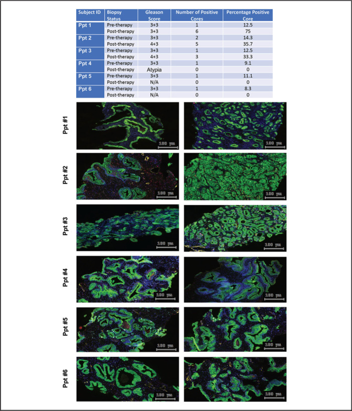
Figure 2.

Histopathological Changes of Prostate Tissue Associated with Noni Treatment. The slides were digitized on the high-resolution TissueFAXS 200 scanner system (Tissuegnostics, Vienna, Austria). Right column, pre-therapy biopsy and left column post-therapy biopsy. 20x magnification immunofluorescence images of prostate tissues show staining with DAPI (blue), CD31 (angiogenesis marker, yellow), Ki67 (proliferation marker, sky blue), cleaved caspase 3 (apoptosis marker, red), and CK8/18 (epithelial marker, green)
