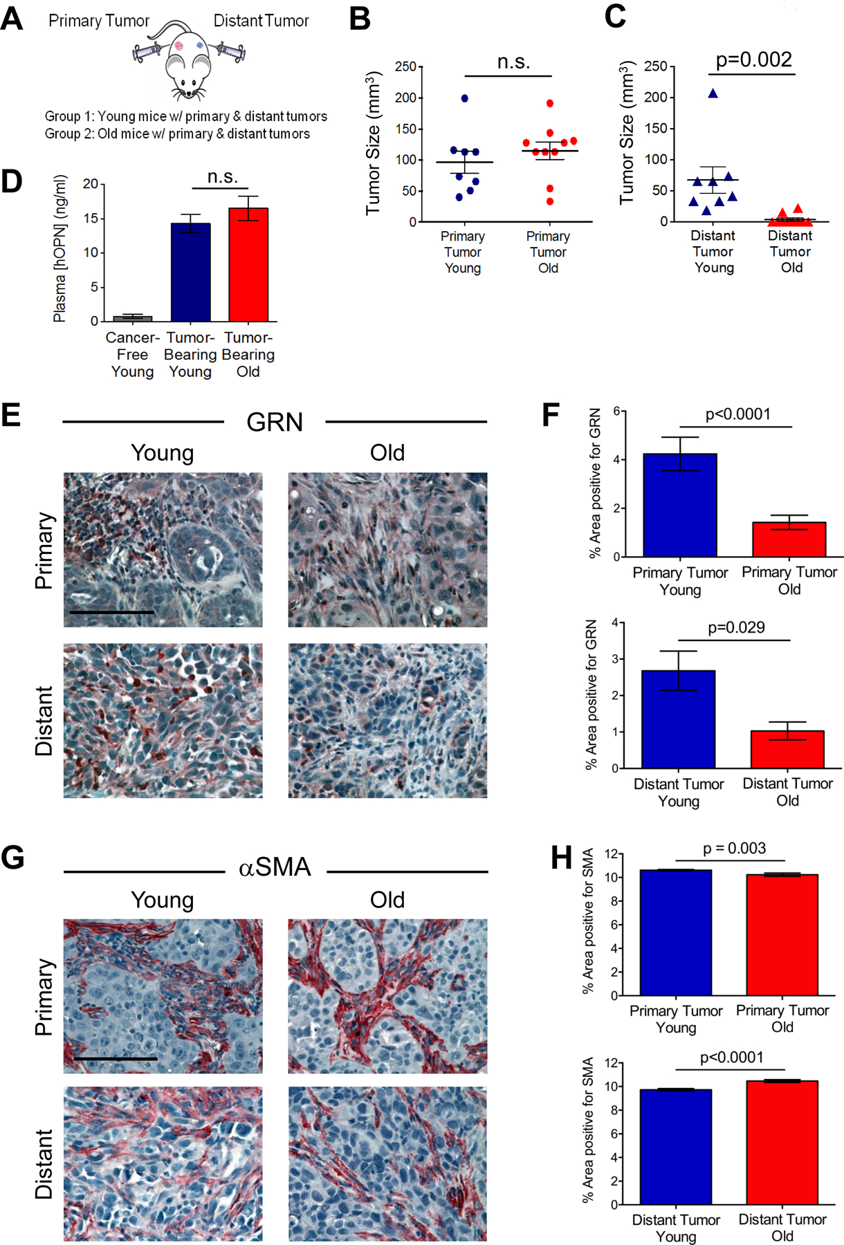
Figure 2. Systemic promotion of tumor progression is attenuated in old mice.

(A) Experimental design to test cancer progression in young and old mice. Distant HMLER-HR tumor cells were injected into young and old mice bearing primary BPLER tumors. Mice were sacrificed at various time points in order to obtain comparably sized BPLER primary tumors between young and old cohorts. (B) Size of primary BPLER tumors from young (n=8) and old (n=10) mice. (C) Size of distant HMLER-HR tumors from young and old mice bearing primary BPLER tumors. (D) Plasma levels of human tumor-derived OPN (hOPN) in young and old mice and cancer-free young mice as a control (n=6 per cohort). (E) Representative images of GRN (red) stained primary tumors (top) and distant tumors (bottom) from young and old mice. Cell nuclei counterstained with hematoxylin (blue); scale bar=100 μm. (F) Quantification of GRN-positive area as a percentage of total area (from E); n=22 images primary young; n=38 images primary old; n=28 images distant young; n=12 images distant old. (G) Representative images of αSMA (red) stained primary tumors (top) and distant tumors (bottom) from young and old mice. Cell nuclei counterstained with hematoxylin (blue); scale bar=100 μm. (H) Quantification of αSMA-positive area as a percentage of total area (from g); n=81 images primary young; n=57 images primary old; n=41 images distant young; n=21 images distant old. Data represent mean ± SEM and analyzed using Student’s t-test for significance. n.s. not significant. See also Figures S1–2.
