. 2021 Aug 31;146(20):6084–6101. doi: 10.1039/d1an00865j

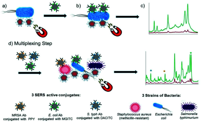
Fig. 4. Schematic illustrating the single-plex and multiplex detection assay. Assay format: (a) lectin (Con A) functionalised silver coated magnetic nanoparticles (Ag@MNPs) will bind to bacteria and the presence of the magnet will allow for magnetic separation of the bacteria from the sample matrix (b) SERS active silver nanoparticles (AgNPs) functionalised with a biorecognition molecule (antibody; Ab) and a unique SERS reporter are added. The mixture is shaken for 30 min before applying a magnet for a further 30 min and allowing the sample to collect. Any unbound matrix is gently removed, and the sample subsequently re-suspended in dH2O (c) The sample is then interrogated with a 532 nm laser beam and SERS signal obtained (green spectrum). When no target is present the functionalised AgNPs will be washed away, thus they will not bind to bacteria, so a minimum SERS signal obtained (red spectrum). (d) Multiplexing step: 3× AgNP conjugates each functionalised with a different Raman reporter and an antibody (which is specific for a bacterial pathogen) are added together with 3 bacterial pathogens and Con A (which binds to all three bacteria) functionalised Ag@MNPs. In the same way as the single-plex systems magnetic separation allows for the samples to be concentrated and analysed via a 532 nm laser. A SERS spectrum is obtained which contains characteristic peaks from the three Raman reporters and thus can be used to confirm if the targets are present. The image is reprinted from Kearns et al.,90 Copyright (2017), with permission from American Chemical Society.

Fig. 4