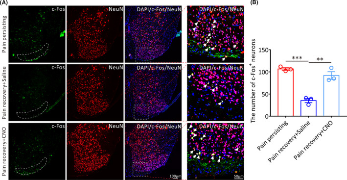
FIGURE 6.

The effect of chemogenetic activation of RVM GPER‐positive neurons on the activation of lumbar spinal dorsal horn neurons in pain recovery mice. (A) Representative immunofluorescence images showing the distribution of c‐Fos+ neurons in the spinal dorsal horn of the three groups mice on 15 days after incision surgery. The white arrows show the c‐Fos+/NeuN+ neurons. Green: c‐Fos+ neurons. Red: NeuN+ neurons. All scale bar, 100 μm or 50 μm. (B) Statistical analysis of the number of c‐Fos+ neurons in the lumbar spinal dorsal horn of three groups mice (n = 3 in each group, each mouse was chosen 10 slices, **p < 0.01, ***p < 0.001, one‐way ANOVA)
