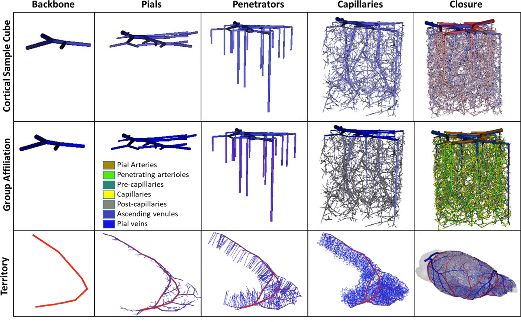
Figure 7.

Visualization of five stages of synthesizing a complete cortical microcirculatory network in mouse. Top row) shows the gradual evolution of small cortical subsection of similar size to E1.1. Second row) shows color coding of anatomical group labels used during the generation of the sVAN. Bottom row) illustrates stages of growing the LACA territory during the construction of the mouse hemisphere. During each stage of growth, new segments are only allowed to connected to specified anatomical groups as outlined in Table 2. The stages show (from left to right): the backbone forms the initial structure onto which the pial network is grown. The penetrating vessel terminals are then generated perpendicular to the pial surface and attached to the exposed pial terminals. Next, the capillaries (precapillary arterioles, pre-capillaries, capillaries, post-capillaries, and post-capillary venules) fill the space until the desired number of vessels is reached. In the capillary stage, it is possible to relax the volume optimization principle to place the optimal bifurcation point without rigorous optimization. In the final stage, closure between the arteries and veins connects all open terminals from the arterial tree to the venous tree and all the venous terminals to the arterial tree as described in Section 2.3.
