
FIGURE 2.
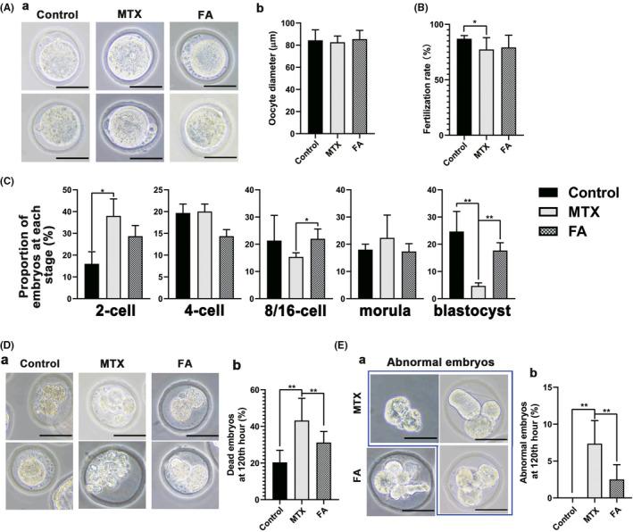
Oocytes from the methotrexate (MTX) group mouse exhibited lower in vitro fertilization (IVF) success rate and abnormal embryo development, which was recovered by folic acid (FA). (A) (a) Representative images of oocytes collected via superovulation after removing cumulus cells. (b) No significant differences were observed in the diameter of oocytes among the three groups. (B) The MTX group exhibited a significantly reduced fertilization rate. FA treatment could not significantly restore the fertilization rate of the oocyte. (C) The percentage of embryos at different stages. The proportion of blastocysts was significantly lower than that of the control group, which was restored after FA treatment. (D) (a) The morphology of dead embryo in each group, the inner cell mass of the dead embryo was fragmented. (b) The statistical chart showed that the proportion of dead embryos in the group was significantly higher than that in the FA treatment group and the control group. (E) (a) Representative images show aberrant embryos from MTX and FA treatment groups including embryos without zona pellucida or with atypical inner cell mass. (b) The statistical chart showed the proportion of aberrant embryos in the MTX group 120 h after IVF, which was much higher than that of the control and FA groups (*p < 0.05, **p < 0.01, Student’s t‐test, n = 6; Scale bars: 50 μm)