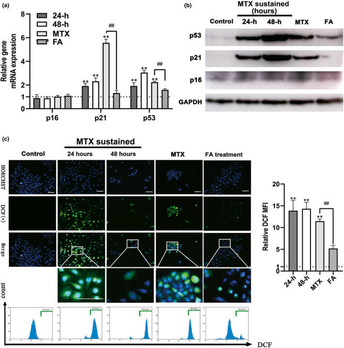
FIGURE 4.

Methotrexate (MTX) caused the OS and increased p53/p21 expression of human granulosa cells (GCs; KGN cells), which can be released by folic acid (FA) antagonism. (A) Detecting the expression of p16, p21, and p53 genes using real time polymerase chain reaction (PCR). MTX upregulated the expression of p21 and p53 but not p16 genes in the KGN cells, p21 and p53 genes maintained the expression at a high level at 72 h after MTX withdrawal. In contrast, the expression of the genes reduced significantly after 72 h of incubation in FA‐medium. (B) Detecting the expression of p16, p21, and p53 genes using Western blot. (C) Detection of OS of KGN cells in MTX, FA treatment, and control group. The reactive oxygen species (ROS)‐positive cells in the green box show the nucleus with blue fluorescence and cytoplasm with green fluorescence. Relative DCF MFI shows quantitative analysis of ROS expression of KGN cells in each group. OS of GCs could be detected as early as 24 h after MTX treatment (MTX sustained group) and still be detected 72 h after MTX withdrawal (MTX group). The addition of FA could eliminate almost all the reactive oxygen species. (Symbol * indicates a difference between MTX processed groups and control group, **p < 0.01; Symbol # indicates a difference between MTX and FA treatment groups, # p < 0.05, ## p < 0.01. Student’s t‐test; Scale bars: 50 μm)
