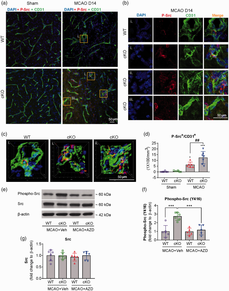
Figure 1.
Src kinase is activated in the peri-infarct regions of EC-targeted miR-15a/16-1 cKO mouse brains after cerebral ischemia. EC-miR-15a/16-1 cKO mice and WT littermate controls were subjected to 1 h MCAO followed by 14d reperfusion. Vehicle (Veh) or AZD0530 (AZD, 20 mg/kg) was administered daily to both genotypes 3–14d after MCAO by oral gavage. CD31 (green), phosphorylated-Src (Y416) (red), and DAPI (blue) triple-immunostaining was utilized to determine the co-localization of cerebral microvessels and activated Src kinase in the peri-infarct regions of the ischemic brains. Yellow boxes indicated areas in (a) that were enlarged in (b) and partly 3D reconstructed in (c). DAPI, 4′,6-diamidino-2-phenylindole. (a–d) Representative images (a–c) and quantitative analysis (d) demonstrated enhanced phosphorylated-Src signals co-localized with cerebral microvessels in the peri-infarcted areas of miR-15a/16-1 cKO mice brains, compared to WT controls. There was almost no positive phosphorylated-Src signals in the sham controls. Data are expressed as the mean ± SD. n = 6–7/group for d. *p < 0.05, ***p < 0.001 between sham and MCAO groups for each genotype; ##p < 0.01 as indicated. Total proteins were also isolated from the ipsilateral cortex of ischemic brains after 14d reperfusion, and western blotting was carried out to detect the protein expression of phosphorylated-Src and total Src. (e,f) Representative western blotting images (e) and quantitative analysis (f) indicated endothelial-selective deletion of the miR-15a/16-1 cluster upregulated phosphorylated-Src (Y416), whereas AZD0530 effectively downregulated the phosphorylation of Src, without significantly altering the expression of total Src (g). Data are expressed as the mean ± SD. n = 5-6/group for f and g. *p < 0.05, **p < 0.01 as indicated. Statistical analysis was performed by one-way ANOVA followed by Bonferroni's multiple comparison tests.

