. 2021 Sep 15;10:e69883. doi: 10.7554/eLife.69883

Figure 1. The endosomal localization of Vps55 requires Mvp1.

(A) Schematic of SNX-BAR proteins in yeast. (B) Model of endosomal recycling pathways in yeast. (C) Schematic of Vps55 and OB-RGRP. (D) Vps55-GFP localization. The mCherry-Pep12 serves as an endosomal marker. (E) Vps55-GFP localization in wild-type (WT), vps35Δ (retromer mutant), snx4Δ (Snx4 complex mutant), and mvp1Δ. (F) Quantification of Vps55-GFP localization from three independent experiments. N = >30 cells. Scale bar: 1 µm.

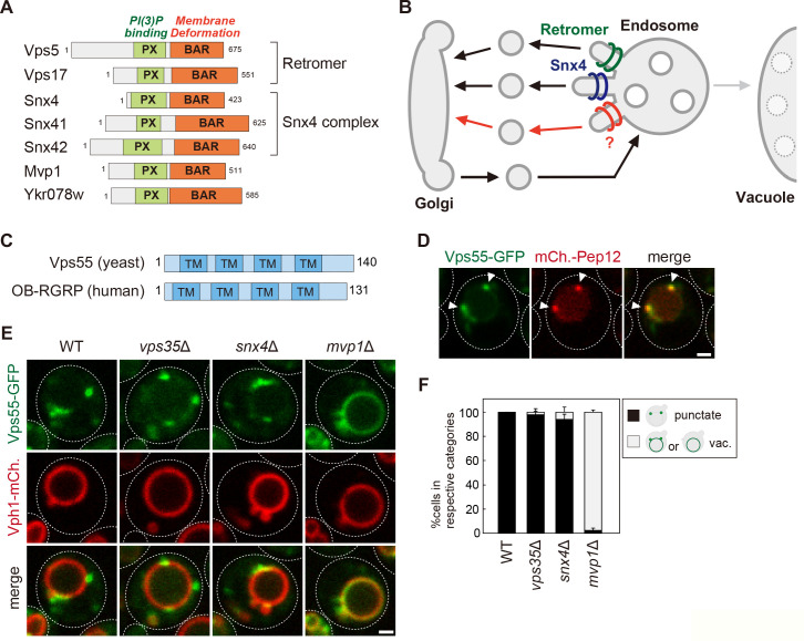
Figure 1—source data 1. Source data associated with Figure 1F.
Figure 1—source data 2. The list of localization altered in retromer mutants. The list of endosomal transmembrane proteins whose localization was examined in vps35Δ cells.

Figure 1.

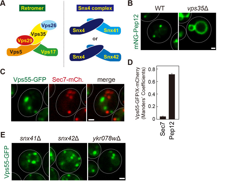
Figure 1—figure supplement 1. The localization of endosomal membrane proteins.

Figure 1—figure supplement 1.

(A) Schematic of retromer and Snx4 complexes. (B) The localization of mNeonGreen-Pep12 in wild-type (WT) and vps35Δ cells. (C) Vps55-GFP localization with Sec7-mCherry serving as a marker for the trans-Golgi. (D) Manders’ coefficients of Vps55-GFP with Sec7-mCherry or mCherry-Pep12 from C and Figure 1D. (E) Vps55-GFP localization in snx41Δ, snx42Δ, and ykr078wΔ cells. Scale bar: 1 µm.
Figure 1—figure supplement 1—source data 1. Source data associated with Figure 1—figure supplement 1D.