. 2021 Sep 15;10:e69883. doi: 10.7554/eLife.69883

Figure 5. Mvp1 mainly mediates retromer-independent endosomal recycling.

(A) Localization of Vps10-GFP in wild-type (WT), vps35Δ, and mvp1Δ cells. (B) Quantification of Vps10-GFP, Kex2-GFP, and GFP-Neo1 localization from A and Figure 5—figure supplement 1E,F, respectively. N = >30 cells from three independent experiments. (C) Schematic for immunoisolation of Vps55-FLAG-containing structures. (D) The immunoisolation of Vps55-containing vesicles. Vps55-FLAG-containing structures were immunoisolated from sec18ts mutants incubated at 37°C for 1 hr, and the isolated structures were analyzed by immunblotting using antibodies against FLAG, Vps10 (retromer cargo), Vps21 (endosome), Pho8 (vacuole), and glucose-6-phosphate dehydrogenase (G6PDH) (cytoplasm). (E) Electron microscopy (EM) analysis of the isolated Vps55-FLAG-containing structures from D. (F) Live-cell imaging analysis of Vps55-mNeonGreen and Vps10-mCherry. (G) Cell growth in vps35Δ snx4Δ mvp1Δ triple mutants. Cells lacking Vps35 as well as Snx4 and Mvp1 were grown at 26°C and 37°C. (H) Model of retromer-, Snx4-, and Mvp1-mediated recycling. (I) Nhx1 localization in SNX-BAR mutants. (J) Quantitation of Nhx1-GFP localization, from I and Figure 5—figure supplement 1M. N = >30 cells from three independent experiments. Scale bar: 1 µm.

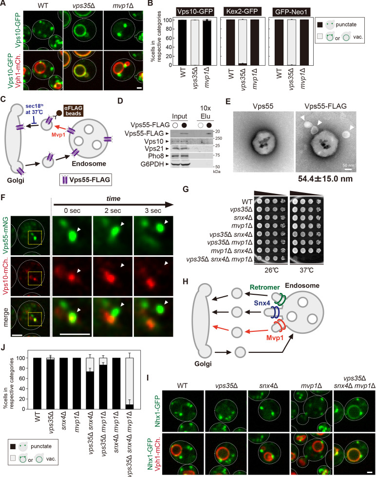
Figure 5—source data 1. Source data associated with Figure 5B.
Figure 5—source data 2. Uncropped gel images of Figure 5D.
Figure 5—source data 3. Source data associated with Figure 5E.
Figure 5—source data 4. Source data associated with Figure 5J.

Figure 5.

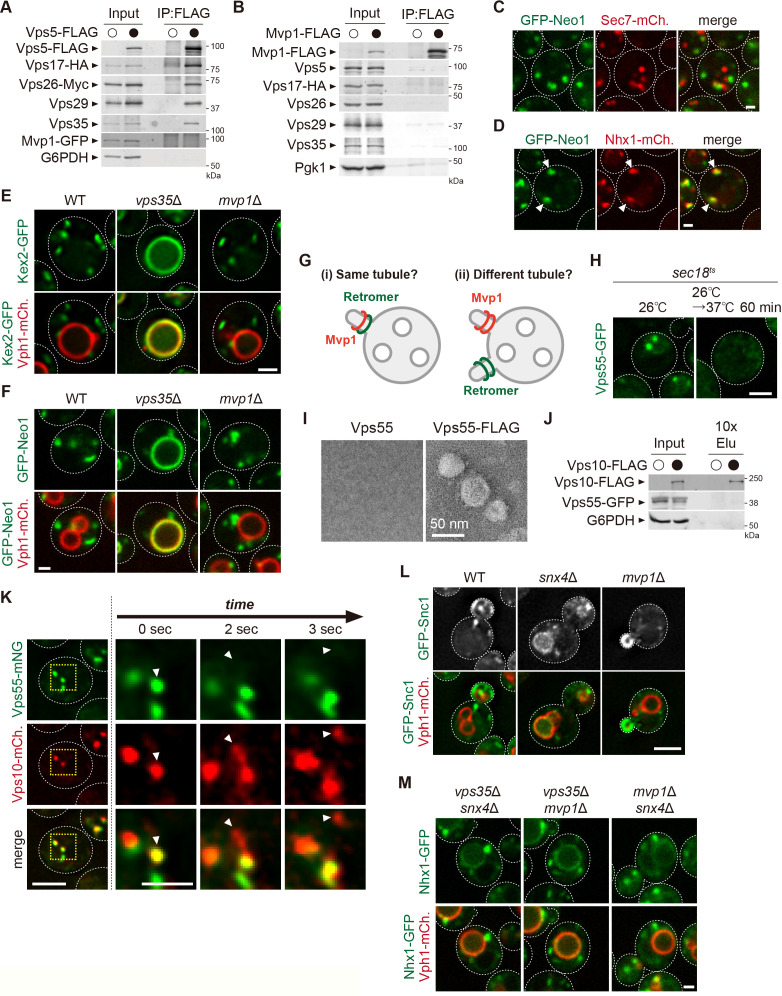
Figure 5—figure supplement 1. The analysis of the retromer pathway in Mvp1 mutants.

Figure 5—figure supplement 1.

(A) The association of Mvp1 with the retromer complex. Vps5-FLAG was immunoprecipitated (IP) from cells expressing Vps17-HA, Vps26-Myc, and Mvp1-GFP, and the IP products were analyzed by immunoblotting using antibodies against FLAG, hemagglutinin (HA), Myc, Vps29, Vps35, green fluorescent protein (GFP), and glucose-6-phosphate dehydrogenase (G6PDH). (B) Mvp1 binding to the retromer subunits. Mvp1-FLAG was immunoprecipitated from cells expressing Vps17-HA, and the IP products were analyzed by immunoblotting using antibodies against Vps5, HA, Vps26, Vps29, Vps35, and G6PDH. (C and D) Localization of GFP-Neo1 with Sec7-mCherry as a marker for the trans-Golgi (C) and Nhx1-mCherry as a marker for the endosome (D). (E and F) Localization of Kex2-GFP (E) and GFP-Neo1 (F) in wild-type (WT), vps35Δ, and mvp1Δ cells. (G) Hypothesis of Mvp1-mediated recycling. (H) Vps55-GFP localization in sec18ts mutants at 26°C or 37°C for 60 min. (I) Electron microscopy (EM) analysis of the isolated Vps55-FLAG-containing structures. Vps55-FLAG-containing structures were immunoisolated from sec18ts mutants incubated at 37°C for 1 hr, and then eluated using FLAG peptides. Eluated products were analyzed by negative-stain EM. (J) Immunoisolation of Vps10-containing vesicles. Vps10-FLAG-containing structures were immunoisolated from sec18ts mutants incubated at 37°C for 1 hr, and the isolated structures were analyzed by immunblotting using antibodies against FLAG, GFP, and G6PDH. (K) Live-cell imaging of Vps55-mNeonGreen and Vps10-mCherry. (L) GFP-Snc1 localization in WT, snx4Δ, and mvp1Δ cells. (M) Nhx1 localization in vps35Δ snx4Δ, vps35Δ mvp1Δ, and snx4Δ mvp1Δ cells. Scale bar: 1 µm.
Figure 5—figure supplement 1—source data 1. Uncropped gel images of Figure 5—figure supplement 1A.
Figure 5—figure supplement 1—source data 2. Uncropped gel images of Figure 5—figure supplement 1B.
Figure 5—figure supplement 1—source data 3. Uncropped gel images of Figure 5—figure supplement 1J.