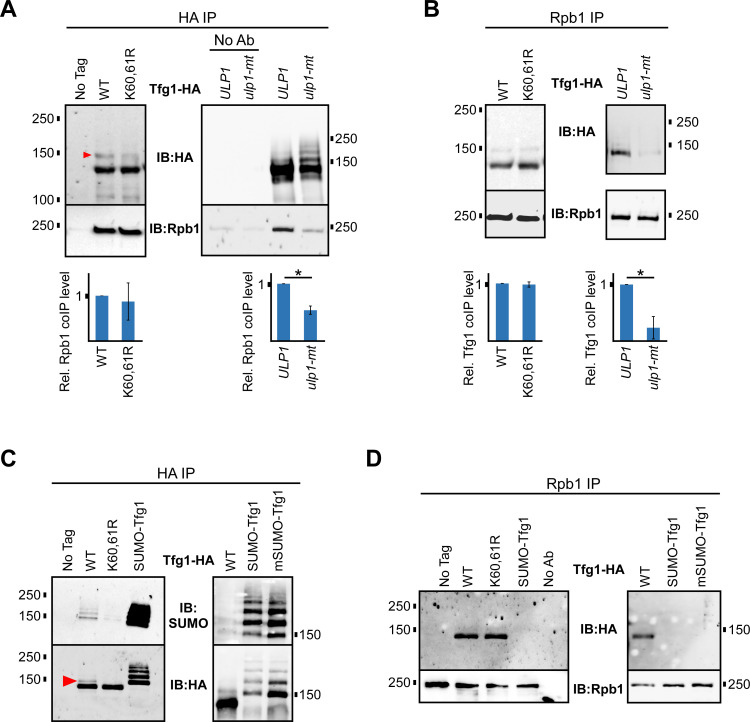
Fig 5. Elevated Tfg1 sumoylation reduces its interaction with RNAPII.
(A) Tfg1-HA and Tfg1-K60,61R-HA strains, and strains expressing Tfg1-HA in the ulp1-mt (I615N) or ULP1 parental backgrounds, were used for co-IP experiments. HA IPs, and no antibody (No Ab) controls, were analyzed by HA and Rpb1 (8WG16 antibody) immunoblots. Relative levels of co-IPed Rpb1, normalized to Tfg1-HA IP levels, were determined by densitometry and average values from at least three experiments are shown. Student’s t-tests were performed and paired values that are statistically different (p value < 0.05) are indicated with an asterisk. (B) Using the same strains described in A, Rpb1 IP was performed, followed by HA and Rpb1 immunoblots. Quantification of levels of co-IPed Tfg1-HA, normalized to IPed Rpb1 levels, was performed as in A. (C) Strains expressing Tfg1-HA with an N-terminal fusion to the yeast SUMO peptide (SUMO-Tfg1) or to a mutant form of the SUMO peptide in which all Lys residues are replaced with Ala (mSUMO-Tfg1), were analyzed alongside strains expressing WT and K60,61R forms of Tfg1-HA in a HA IPs, followed by SUMO and HA immunoblots. (D) Strains expressing WT, K60,61R, SUMO-Tfg1, or mSUMO-Tfg1 forms of Tfg1-HA were used for Rpb1 IP followed by immunoblot analysis with HA and Rpb1 antibodies.

