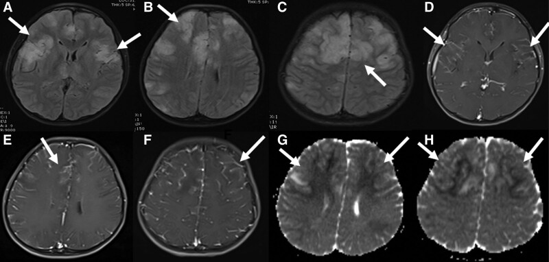
FIGURE 1.
Brain MRI of a 9-year-old boy at presentation. In the fluid-attenuated inversion recovery sequences, pathologic signal changes were observed in cortical and subcortical areas in bilateral frontoparietal regions (A–C). These lesions showed contrast enhancement after contrast enhancement (D–F) and marked diffusion restriction in apparent diffusion coefficient mapping in diffusion-weighted sequences (G and H).

