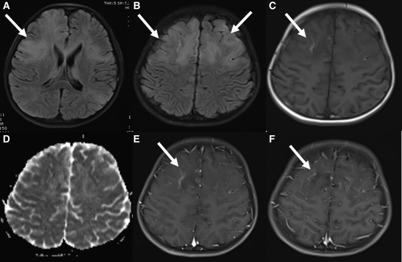
FIGURE 2.
Brain MRI of a 9-year-old boy on day 9. The lesions continued in fluid-attenuated inversion recovery sequences (A and B) and cortical linear hyperintensities compatible with laminar necrosis occurred in T1A sequences (C). It was found that the restriction disappeared in the diffusion-weighted sequences (D) and contrast enhancement continued in the frontoparietal regions after contrast agent administration (E and F) and necrotic areas began to occur in these regions.

