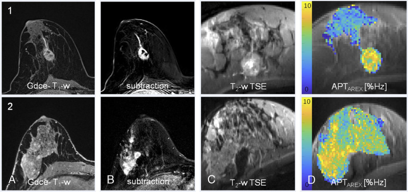
FIGURE 8.

Relaxation-compensated APT CEST-MRI at 7 T and clinical MR mammography. Patient 1: high grade/patient 2: intermediate grade intraductal breast cancer of no special type. In both patients, a strong gadolinium enhancement can be observed at clinical MRI: gadolinium-enhanced (Gdce), fat-saturated T1-weighted MRI after administration of a standard dose (0.1 mmol/kg body weight) of gadobutrol (TR, 28 milliseconds; TE, 4.76 milliseconds; slice thickness, 1.1 mm; flip angle, 25; field of view, 360; matrix, 320) and subtraction MRI. In addition, T2-weighted TSE (1c, 2c) and the APTAREX contrasts at 7 T are shown. All breast cancers could be clearly identified on the APTAREX contrast. APT signal hyperintensities showed a distinct morphological correlation with the contrast-enhanced MR images. Reproduced with permission from Elsevier, Loi et al.335
