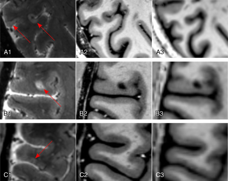
FIGURE 2.

Cortical MS lesions. Ultra-high-field imaging at 7 T allows for detection of all types of cortical MS lesions (red arrows; A1–A3, subpial lesions; B1–B3, leukocortical lesion; C1–C3, intracortical lesion), exemplified using a T2*-weighted gradient echo sequence (T2*w GRE; spatial resolution, 0.5 mm isotropic) for the detection of a subpial lesion (A1), a leukocortical lesion (B1), and an intracortical lesion (C1). T1-weighted sequences, such as the magnetization prepared 2 rapid acquisition gradient echo (MP2RAGE), are also sensitive to cortical MS lesions at 7 T (A2, B2, C2; spatial resolution, 0.5 mm isotropic; average of 4 acquisitions) compared with 3 T (A3, B3, C3; spatial resolution, 1 mm isotropic; single acquisition).
