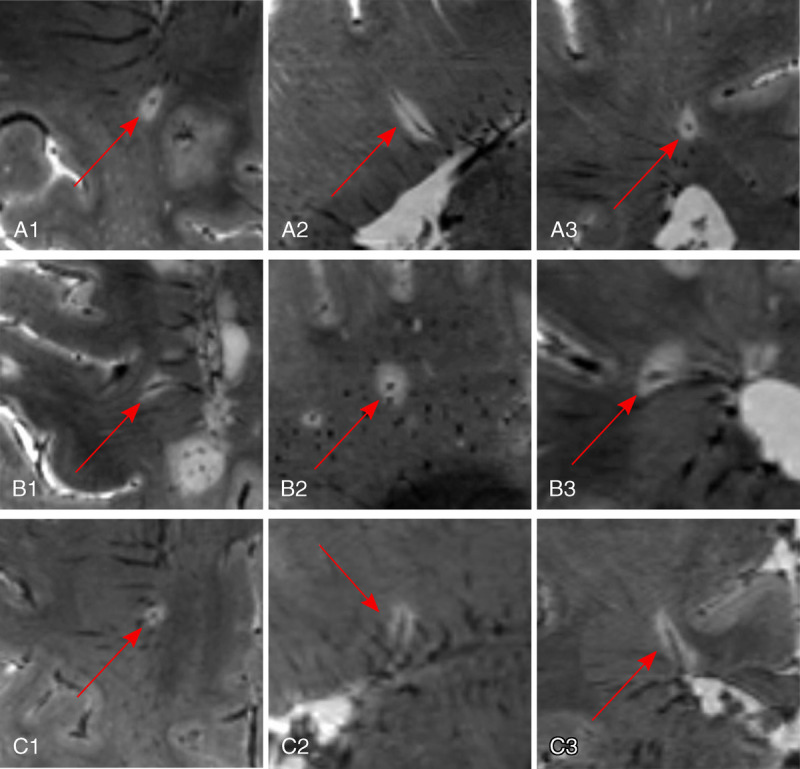
FIGURE 3.

Central vein sign in MS. Ultra-high-field imaging at 7 T yields conspicuous central veins within MS lesions even when small in diameter, exemplified in 3 cases (A–C) by using a multiecho T2*w gradient echo sequence (spatial resolution, 0.5 mm isotropic). The veins are centrally located within the lesion in all 3 planes (A1, B1, C1: axial plane; A2, B2, C2: sagittal plane; A3, B3, C3: coronal plane).
