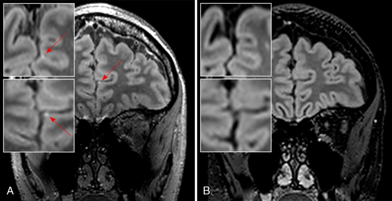
FIGURE 4.

Leptomeningeal enhancement in MS. Ultra-high-field imaging at 7 T is able to sensitively detect foci of leptomeningeal enhancement (LME, red arrow) as demonstrated in this postgadolinium T2-weighted fluid-attenuated inversion recovery (T2-FLAIR; spatial resolution, 0.7 mm isotropic) sequence from a progressive MS patient with interhemispheric LME (A, with magnified axial [top] and coronal [bottom] images), which is not detected on postgadolinium T2-FLAIR images at 3 T (B, with magnified axial [top] and coronal [bottom] images). Both T2-FLAIR images were acquired ≈10 minutes after contrast medium administration.
