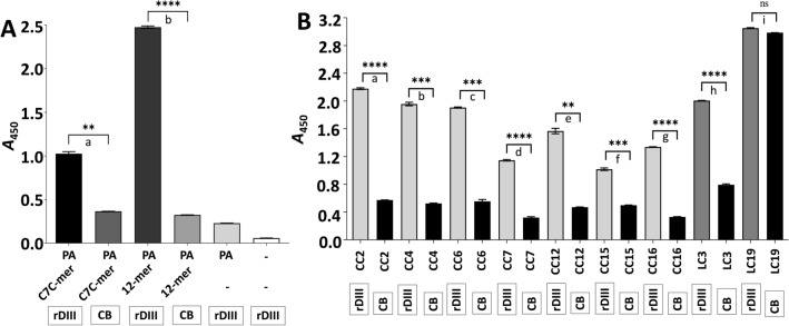
Figure 1.
Confirmation of the interaction between rDIII and amplified phages using semi-quantitative ELISA. (A) Phage ELISA demonstrating the interaction between rDIII and C7C-mer and 12-mer phages amplified from the last round of biopanning. (B) Phage ELISA demonstrating the interaction of individual C7C-mer and 12-mer phage clones to rDIII. Framed reagents were coated into microtiter wells. Data present mean of triplicates with ± S.D. Asterisks indicate statistical significance. A statistical significance difference (P < 0.01, two-tailed P-value) was calculated with paired t-test. Statistics was performed with statistics tool of GraphPad Prism v8.4.3. In Panel (A)—a: P = 0.0013; b: P < 0.0001. In Panel (B)—a: P < 0.0001; b: P = 0.0002; c: P = 0.0007; d: P < 0.0001; e: P = 0.0014; f: P = 0.0010; g: P < 0.0001; h: P < 0.0001; i: P = 0.037 (ns). A – Absorbance; CB – coating buffer; rDIII – recombinant domain III; ns – non-significant; PA – primary antibody, CC – phage clone carrying 7-mer cyclic peptide; LC – phage clone carrying 12-mer linear peptide.

