Figure 5.
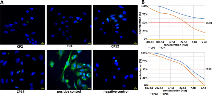
Blocking of the adhesion of rDIII on cultured endothelial cells and neutralization assay. (A) Blocking of the adhesion of rDIII on endothelial cells. Nuclei are stained with DAPI. The assay was performed in triplicates. Positive control – rDIII was incubated with the cells. Negative control – rDIII was excluded from the assay. CP – 7-mer cyclic peptide. (B) Assessment of the ability of peptide to block infection in cultured cells (neutralization assay). Neutralization of the infection of virus like particle carrying Fluc gene by CP2, CP4, CP12 and CP16. The amount of VLP entering the target cells was calculated by detecting the expression of luciferase, which then used to measure the neutralizing ability of the peptides, expressed in half maximal effective concentration (EC50). Neutralizing capacity of each CP was observed as follows: CP2 – EC50 > 7290 (17.3 ng/ml, 2.43 nM), CP4 – EC50 1144 (119.4 ng/ml, 17 nM), CP12 – EC50 1811 (73.3 ng/ml, 10.4 nM) and CP16 – EC50 1270 (104.7 ng/ml, 14.8 nM).

