Figure 3.
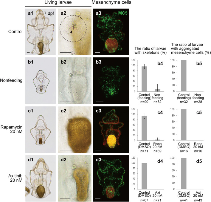
Observations of skeleton formation in the experimental starfish larvae. Morphology and skeleton formation were observed in control larvae (a1–a3), nonfeeding larvae (b1–b5), rapamycin (rapa)-treated larvae (c1–c5), and axitinib (axi)-treated larvae (d1–d5) at 7 dpf. (a1–d1, a2–d2) Living larvae. In the panel a2, the arrowhead indicates an adult skeletal rudiment and the dotted circle indicates aggregated mesenchyme cells. (a3–d3) Fluorescence images of larvae examined by immunohistochemistry using the mesenchyme-specific marker MC5. The green signal shows MC5 expression, whereas the red signal shows Chaetoceros calcitrans in the stomach or autofluorescence. At 7 pdf, the ratio of larvae with adult skeletons (b4–d4) and that of larvae with aggregated mesenchyme cells in the posterior dorsal region (b5–d5) were evaluated. Feeding larvae and DMSO-treated larvae were used as controls for nonfeeding larvae and inhibitor-treated larvae, respectively. Scale bars: 50 μm. M: mol/L.

