Figure 1.
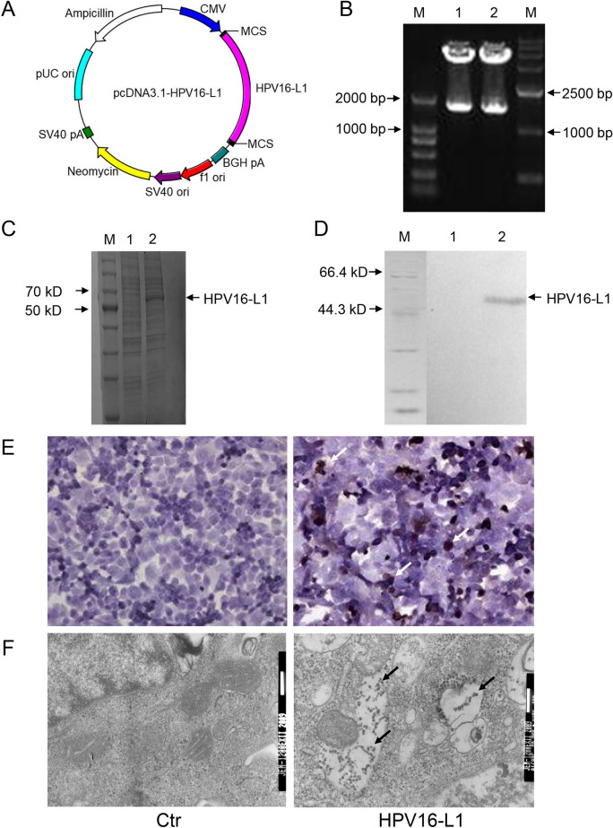
The pcDNA3.1-HPV16-L1 plasmid was successfully constructed and HPV16-L1 was identified to express in BHK cells. (A) The map of pcDNA3.1-HPV16-L1. (B) Identification of pcDNA3.1-HPV16-L1 by restriction enzyme digestion. (M1: DL2 000; lane 12: plasmid digested by HindIII and KpnI; M2: DL15000). (C) SDS PAGE analysis of HPV16-L1 protein expressed in BHK cells (M: protein marker, lane 1: control, lane 2: expressed protein). (D) Western blotting analysis of HPV16-L1 protein expressed in BHK cells (M: protein marker; lane 1: control; lane 2: expressed protein). (E) Immunocytochemistry analysis of HPV16-L1 protein expressed in BHK(× 400). (F) Electron microscropy detection of HPV16-L1 expressed in BHK cells.

