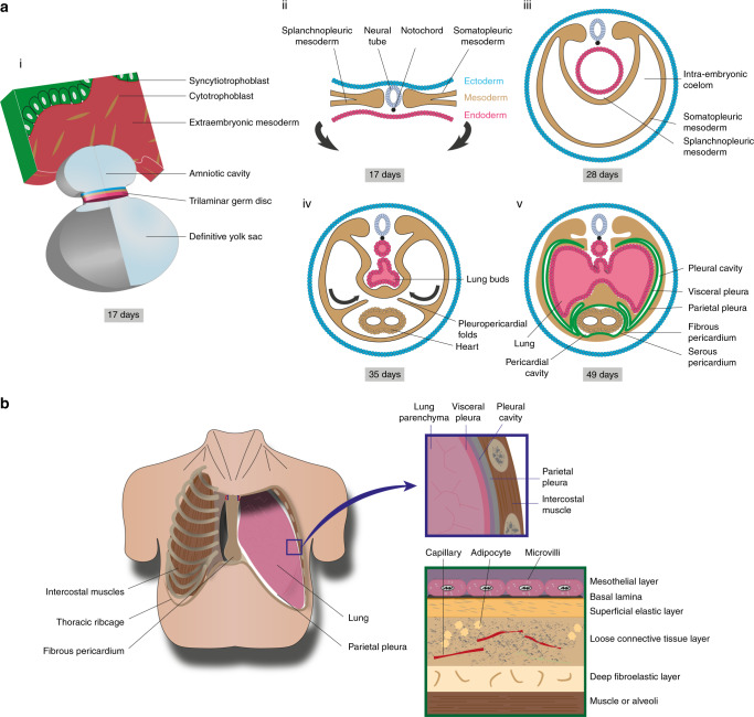
Fig. 4. Pleural anatomy.
a Embryonic pleural development. (i) Representation of the trilaminar germ disc following gastrulation together with the amniotic cavity and definitive yolk sac. (ii) During gastrulation, the mesoderm layer forms and the lateral plate mesoderm subdivides into somatic and splanchnic mesoderm. (iii) Following lateral flexion, the intra-embryonic coelom is lined by somatic and splanchnic mesoderm. (iv) The pleuropericardial folds extend to the midline and fuse with the ventral surface of foregut mesoderm, forming the primitive pleural cavities. (v) The somatic and splanchnic mesoderm is lined by parietal and visceral pleura respectively, which are contiguous with each other at the level of the hilum. b Anatomy of the thorax showing the relationship between the lungs, thoracic ribcage and pleura. The pleural cavity is defined by the space between adjacent visceral and parietal pleura. The pleura can be subdivided into five layers: mesothelial cells, basal lamina, superficial elastic layer, connective tissue layer and the deep fibroelastic layer. The deep layer is tightly adhered to the underlying structures e.g. muscle, rib or lung parenchyme.

