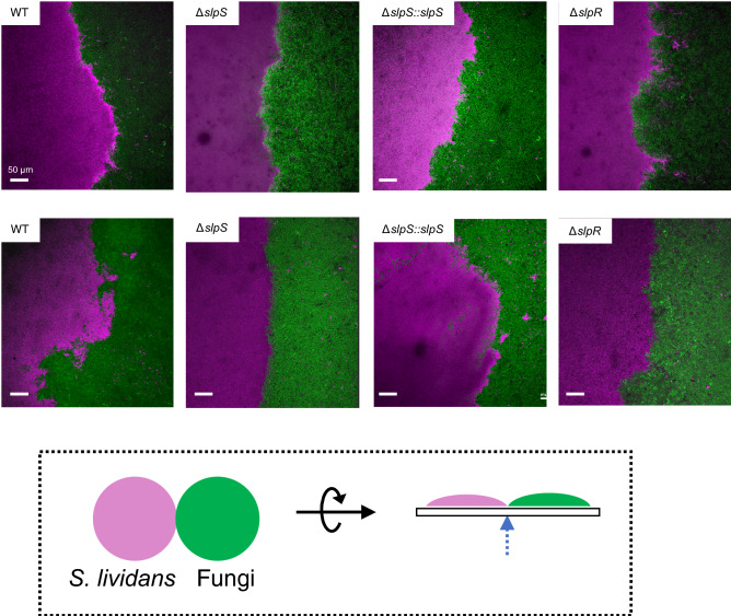
Figure 4.
Confocal laser scanning microscopy (CLSM) of the colony boundaries between S. lividans and fungi. Colony boundaries between S. lividans and fungi were observed by CLSM. (A) S. lividans strains were co-cultured with S. pombe (top) and S. cerevisiae (bottom) on a cellophane, and the bottom of the colony boundary was then observed by CLSM. A dotted arrow in the bottom panel indicates the observed region between the colonies. S. lividans and fungi were visualized using Syto59 dye staining (magenta) and GFP expression (green), respectively. Scale bars, 50 μm.

